The Neurological Exam Explained
We have all had a thorough once-over by a neurologist, and chances are you see one at least once a year for a checkup and exam. Have you ever obediently walked around on your tippy toes or moved your finger from your nose to your doctor's finger over and over, wondering what it is they are looking for or secretly hoping that this isn’t just a trick to make you look ridiculous?
All these tests and hoops your doctors make you go through give them a good look at how the nervous system is working. But people often ask me what exactly we learn from doing them. I wanted to take the time to elaborate a bit more on each aspect of the exam so you can understand its purpose a little bit better.
MS neurological exam: Testing the cranial nerves
Cranial nerves start in the brain and control various movements and sensations of the face and neck. There are a total of 12 cranial nerves, and MS can affect them in a variety of ways. Neurologists will sometimes test them one by one or focus on the nerves that are most relevant to a specific complaint.1,2
Below is a list of each nerve, what it controls, and how we test it.
1. Olfactory nerve
This controls our sense of smell. We usually do not test this nerve because people can report a change in smell accurately.1
2. Optic nerve
This controls vision, color perception, and visual fields. The optic nerve is commonly damaged by MS, and there are a lot of different ways to test it.1,2
Eye charts help detect changes in vision, such as blurred or double vision. In people who have MS sometimes colors are less vivid in one eye. We can test this by holding up a red square and seeing if it looks like the same shade of red in both eyes. To test peripheral vision, we hold our hands way out to the sides of the person’s face and ask them to tell us how many fingers we are holding up or if they can see which fingers are moving. Finally, we turn off the lights in the room and look at the back of the eye with a bright light.1,2
3. Oculomotor nerve
This nerve constricts the pupils, opens the eyelids, and controls the movement of the eye (extraocular movements). We test this by shining a light in the eyes to see if the pupils constrict properly and by having the person follow our finger as we move it up, down, and side to side.1
[banner class=bnrOneToOneC]
4. Trochlear nerve
This nerve also moves the eye and is specifically responsible for moving the eye down and inward. This is also tested by having the person follow our finger with their eyes.1
5. Trigeminal nerve
This nerve moves the jaw and processes sensory information from the face. Facial pain, called trigeminal neuralgia, is a common MS symptom. To test this nerve we have the person clench their jaw, and we look to see if it is symmetrical. We can also test sensation by touching each side of the face to see if the sensation is the same on each side.1,2
6. Abducens nerve
This is responsible for moving the eyes to the side (away from the nose). Again, we test this by having people follow our fingers with their eyes.1
7. Facial nerve
This nerve controls the movement of the face and our sense of taste. To test it we have people make facial expressions like smiling, squeezing their eyes shut, puffing their cheeks, and raising their eyebrows to see if both sides of the face are symmetrical.1
8. Acoustic/vestibulocochlear nerve
This nerve is responsible for hearing and balance. To test this nerve we can either test the hearing by seeing if the person can hear a soft sound, such as whispering in each ear or by using a tuning fork. A tuning fork is a metal, fork-like instrument. When it is struck it produces audible vibrations, and we can see if one ear hears the vibrations better than the other ear.1
9. Glossopharyngeal nerve
This nerve also plays a role in our sense of taste, and movement of the soft palate. It also controls the gag reflex. It is tested by watching the palate rise with the person’s mouth open. Luckily we don’t force you to gag to test this one!1
10. Vagus nerve
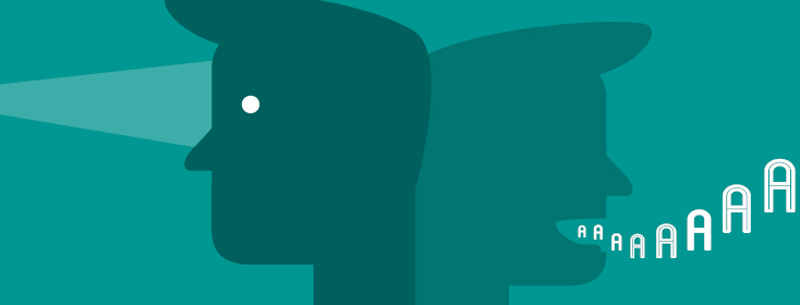
This controls the movement of the palate and throat. To assess this nerve we have the person say “ahh” and watch the movement of the uvula to ensure that it is in the middle and not deviated to the side.1
11. Spinal accessory nerve
This nerve moves the neck muscles. To test its function we have the person move their head from side to side and shrug their shoulders against resistance. To do this, we place our hands on the face and shoulders, and the person has to overcome the resistance of our hand to complete the motion.1
12. Hypoglossal nerve
This nerve controls the movement of the tongue. To test it we have the person stick out their tongue and look to see if it is in the middle. We also look to see if there are any signs of muscle weakness in the tongue. Most people love the opportunity to stick their tongue out at their doctor!1
MS neurological exam: Testing the brain and the spinal cord
There are several ways we test the brain and spinal cord. These tests provide us with a lot of information about how the nervous system is functioning overall and whether there are any disruptions in communication.
Walking
The way a person walks (or their gait) tells us a lot about how well the nervous system is working. We look for muscle weakness, foot drop, imbalance, and speed. We will also have a person walk on their toes, heels, and in a straight line (heel to toe) to check their balance and the strength of different muscle groups.3
Reflexes
These are involuntary movements controlled by the spinal cord. Absent, unequal, or weak reflexes indicate a lesion on the spinal cord. Exaggerated reflexes or clonus (rhythmic muscle movements/oscillations) can also be a result of spinal cord lesions.3
Balance
One of the most common ways of assessing balance is to have the person stand up straight and close their eyes. If they lose their balance with their eyes closed, this is usually a sign of a spinal cord lesion.3
Sensation
Sensory nerves travel from the spinal cord to the brain and can be tested by seeing if a person can tell the difference between a sharp and dull, cold and hot, and if they can feel vibration.3
Coordination
To test coordination we check to see how well a person can do fine movements, such as tapping their fingers together, rapidly moving their hand, and moving their finger back and forth from their nose to the doctor’s finger. Another way to assess coordination is to have the person run the heel of their right foot up and down their left shin, and vice versa. These coordination tests tell us how well a part of the brain called the cerebellum is functioning.3
Strength
We will have the person move their arms and legs against resistance to see how strong they are, and if both sides of the body are equally strong. To do this we have the person push and pull our hands with their arms and legs, and squeeze our fingers.3
Range of motion
If the person has spasticity of the muscles, it will limit how much movement they have in their joints. Moving the joints of the legs and arms helps us to determine how severe muscle spasticity is.3
I hope this helps you understand what it is your doctor is looking for every time you go in for your checkups! Have you ever had any other exam done that left you wondering, "What on earth are they looking for?" Let me know in the comments below!

Join the conversation